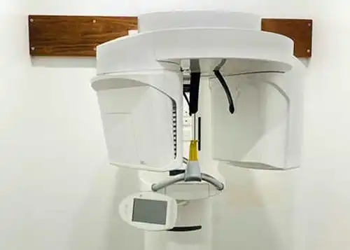
A CBCT, or Cone Beam Computed Tomography, is an advanced type of X-ray that provides dentists with a detailed, 3D view of a patient’s mouth, face, and jaw. Unlike a standard dental X-ray, which only produces a flat, two-dimensional image, a single CBCT scan can capture a comprehensive picture of the teeth, soft tissues, nerve pathways, and bone structure. This makes it an essential tool for situations where a more in-depth look is needed.
Cone Beam CT
CBCT vs. Conventional CT Scans
Cone Beam CT (CBCT) differs from a traditional CT scan in several key ways, making it a more efficient and cost-effective option for dental and craniofacial imaging. While both technologies can produce similar high-quality, 3D images, a CBCT scan offers distinct advantages:
Technology: Unlike a conventional CT scan that uses a fan-shaped X-ray beam and one-dimensional detectors, a CBCT scan utilizes a cone-shaped beam and two-dimensional detectors. This allows it to capture a wider area in a single pass.
Efficiency: CBCT scanners are much faster and can be conveniently used right in a dental office. This saves both time and money.
Precision: By taking multiple angles and views at once, a CBCT scan provides dentists with a more comprehensive evaluation, which leads to more precise treatment planning.
Dental Cone Beam CT Advantages
This type of CT scan gives an accurate view of the bone in detail and is helpful to evaluate diseases of the jaw, teeth, face, and sinuses. Using this technology, your doctor can produce three-dimensional (3-D) images of your teeth, soft tissues, nerve pathways, and bone in a single scan.
Cone Beam CT Has Several Advantages, Including:
Decrease in examination time
Decrease in patient movement
Increased x-ray tube efficiency
The CT scans are painless, non-invasive, and accurate
What Are Some Common Uses Of The Procedure?
By using cone beam CT scans, the dentist can plan a more precise treatment than with conventional dental x-rays. Bone and soft tissue can both be viewed by CT simultaneously, which is one of its major advantages. Dental cone beam CT is commonly in use for for more complex cases that involve:
Planning the surgical removal of impacted teeth
The diagnosis of temporomandibular joint disorder (TMJ)
Placing dental implants accurately
Examination of the jaw, sinuses, nerve canals, and nasal cavity
Detecting, measuring and treating jaw tumors
Understanding the anatomy of the jaw and the orientation of the teeth
Finding the source of pain or pathology
How Does The Procedure Work?
We In a cone beam CT examination, the gantry or C-arm rotates around the head in a complete 360-degree rotation, it creates a 3-D image by capturing multiple images from different perspectives and reconstructing them into one.
X-ray source and detector are mounted on opposite sides of the rotating gantry and rotate simultaneously. A single rotation of the detector can provide up to 200 high-resolution two-dimensional (2-D) images, which combines digitally to produce a 3-D image for your dentist or oral surgeon to review.
How Is The Procedure Performed?
The dentist will position you so that the area of interest falls in the center of the beam. As the x-ray source and detector revolve around you for a 360-degree rotation or less, you will need to remain very still. On average, the entire mouth and dental structures takes about 20 to 40 seconds for a full mouth x-ray.
Dr. Ajay is a member of the Australian Dental Association and Australian Society of Implant Dentistry. Dr. Ajay keeps abreast with the advances in dentistry by attending course and lectures in Australia and around the world. Furthermore, as a practicing dentist, he loves to listen to your questions, concerns and explain all your treatment options. Call (07) 4151 7305 for an appointment